MRI brain Scan
What is an MRI scan?
An MRI scan is a safe, painless way to make detailed images of the brain, nervous system and other soft tissues. It uses strong magnets and radio waves to help us visualize structures and brain activity, without using any radiation.
As a centre of excellence in neurology, we use MRI as one of the most up-to-date technologies for diagnosing neurological conditions, guiding treatment plans, and monitoring progress.

Our technology
SIGNA™ Hero
The center of excellenge Affidea neuraCare, in collaboration with GE HealthCare, introduces the 3T SIGNA™ Hero MRI scanner. This edition brings state-of-the-art imaging technology, combining advancements in MRI with the sophisticated engineering of a modern system. This advanced MRI offers a unique ability to image bones completely free of artifacts using zeroTE – oZTEo technology, reducing the need for examinations with ionizing radiation.
The SIGNA™ Hero performs examinations with maximum comfort for patients as it provides:
- A wide 70 cm bore to minimize discomfort caused by claustrophobia
- Unique AIR™ Coils technology, perfectly adapting to each patient without applying any pressure or creating a sense of restriction
- Advanced AI-powered image generation technology, AIR™ Recon DLTM, enabling ultra-fast, high-quality diagnostic scans while significantly reducing exam time.

AIR™ Coils
The use of conventional rigid coils in MRI technology has long been considered a limitation for both the operator and the patient experience. Traditional MRI coils are usually bulky, heavy, rigid, and uncomfortable, both for patients and healthcare professionals. The new AIR™ coils from GE HealthCare are designed to optimally adapt to patient anatomy thanks to their unique flexibility, resembling a comfortable blanket.
This innovative technology uses a groundbreaking conductive material designed for exceptional flexibility, while each coil is 60% lighter than conventional ones. Flexible enough to wrap around the body with precision, they deliver high-quality images. Their lightweight and unique adaptability minimize physical strain during the exam, significantly improving patient comfort and reducing scan time.
GE HealthCare is revolutionizing MRI with AIR™ Coils, combining patient comfort, image quality, and ease of use.

ΑIR Recon DL
AIR™ Recon DL is a pioneering image reconstruction software based on Deep Learning technology, which can deliver scans with unparalleled accuracy and diagnostic value in half the time compared to traditional MRI systems. The images produced with AIR™ Recon DL are cleaner and sharper than conventional MRI scans, significantly enhancing diagnostic precision for radiologists.
AIR Touch
The SIGNA™ Hero MRI scanner has the ability to automatically adjust the signal acquisition devices (the MRI coils) for each patient, ensuring the optimal exam protocol every time. This means that every scan delivers the best possible results without the operator having to manually intervene, thanks to multiple smart options that either improve image quality or shorten scan time.
AIRx
The SIGNA™ Hero MRI is also equipped with AIRx, a Deep Learning technology that ensures consistent, high-quality image acquisition by automatically identifying the anatomy being scanned and selecting the most suitable imaging parameters, regardless of patient variability or disease. This technology is especially useful for patients undergoing repeat scans, as it provides highly comparable images across time, helping track disease progression more accurately.
*AIRx is particularly compatible with neurological exams of the brain and spine.
SIGNA™ Works
The new SIGNA™Works platform redefines the concept of productivity through its wide range of Core Imaging techniques, offering a series of advanced solutions. With its core applications, SIGNA™Works provides an extensive suite of high-quality imaging tools and features that enable the achievement of optimal results across the full spectrum of MRI applications.
The SIGNA™ Hero MRI scanner comes with SIGNA™Works as a complete and integrated solution. Its added value lies in its ability to continuously upgrade and adapt to the evolving needs of diagnostic centers. The SIGNA™Works platform fully leverages the potential of DirectDigital Imaging (DDI), further enhancing diagnostic capabilities while remaining patient- and user-friendly.
Speed and Simplicity
The powerful gradients of the SIGNA™ Hero, combined with the full set of CV Works acquisition techniques, ensure top-level cardiac imaging.
- Turbo LAVA / LAVA Flex techniques allow faster whole-body imaging with up to 47% shorter breath-holds through multi-phase acquisitions in a single breath-hold, ensuring accurate results from the very first attempt.
- The VIBRANT technique, combined with an eight-channel breast coil, enables high-quality breast imaging and MRI-guided biopsy.
HyperWorks
This new generation of ultra-acceleration tools (HyperSense, HyperCube, HyperBand) uses optimized options to accelerate data acquisition and reduce scan time. Sparse data sampling and adaptive RF are applied for volumetric imaging and fast slice excitation for diffusion studies. The HyperMAVRIC SL technique enables high-quality imaging even in the presence of metallic implants (e.g., hip replacements, orthopedic prostheses, etc.).
Together, the HyperWorks tools can be applied to accelerate a wide range of examinations.
*Metallic implants must always be MRI-compatible.
SilentWorks
GE’s exclusive SilentScan technology dramatically reduces scan noise from 91 dB to as low as 3 dB, below the level of ambient noise in the exam room.
Types of MRI
There are three main types of MRI that we use to create images of the brain:
- MRI – used to create images of the brain tissue and structures
- Functional MRI (fMRI) – shows areas of activity in the brain
- Magnetic resonance angiography (MRA) – used to create images of blood vessels in the brain
Our scanners use 3 Tesla (3T) MRI. The powerful magnetic field allows fast scans that give clear, detailed and accurate images to optimize diagnosis and treatment planning.
Book a consultation
What is an MRI scan used for?
An MRI scan of the brain is often used to diagnose, monitor, and plan treatment for neurological conditions. A specialist neurologist will review your results alongside a range of other tests and scans needed for an accurate and clear understanding of your health, including any next steps in treatment.
These include:
- Brain injury
- Brain and spinal tumors
- Compressed nerves in the spinal cord (myelopathy)
- Vascular problems, such as a bleed, aneurysm or malformed blood vessels (arteriovenous malformations, dural fistulas or cavernomas)
- Brain damage related to epilepsy
- Neurodevelopmental conditions
- Genetic syndromes, such as tuberous sclerosis complex
- Alzheimer’s
- Parkinson’s disease
- Multiple sclerosis (MS) and other inflammatory disorders, such as transverse myelitis
- Myasthenia gravis (a neuromuscular condition)
You may also have a brain MRI for other conditions, such as headaches or migraines, infections, malformations of the brain or spinal cord, or cysts. Although these are not diagnosed through an MRI, the scan helps rule out other possible causes and supports your overall diagnosis and care plan.

Our multidisciplinary team
We surround our patients with specialists who listen, understand, and care. They include some of Europe's most renowned experts – neurologists,
neuropsychologists, neuroradiographers, microbiologists, nurses, nuclear medicine experts and therapeutic specialists in physiology and occupational health.
What to expect during an MRI scan
A brain MRI is a quick, safe and painless scan. It takes around 15 to 20 minutes, but you can expect to be at the clinic for longer depending on any other tests, scans or appointments you may have.
Before your scan
You don’t usually need to do anything to prepare for a brain MRI. Your care team will let you know if there’s anything you need to do.
You’ll complete a safety questionnaire before the scan. Let your neuroradiographer know if you have any implanted devices or are pregnant, as an MRI may not be suitable.
When you arrive at your appointment, you’ll be shown to a private room, and we’ll help you get ready. You don’t usually need to undress for a brain MRI, but you will need to remove any head coverings, jewelry, watches, dentures, hearing aids or wigs.
If your child is having an MRI scan, they may be given a sedative or general anaesthetic to help them feel more relaxed and comfortable. This can also be an option for people who are claustrophobic or have conditions or symptoms preventing stillness.
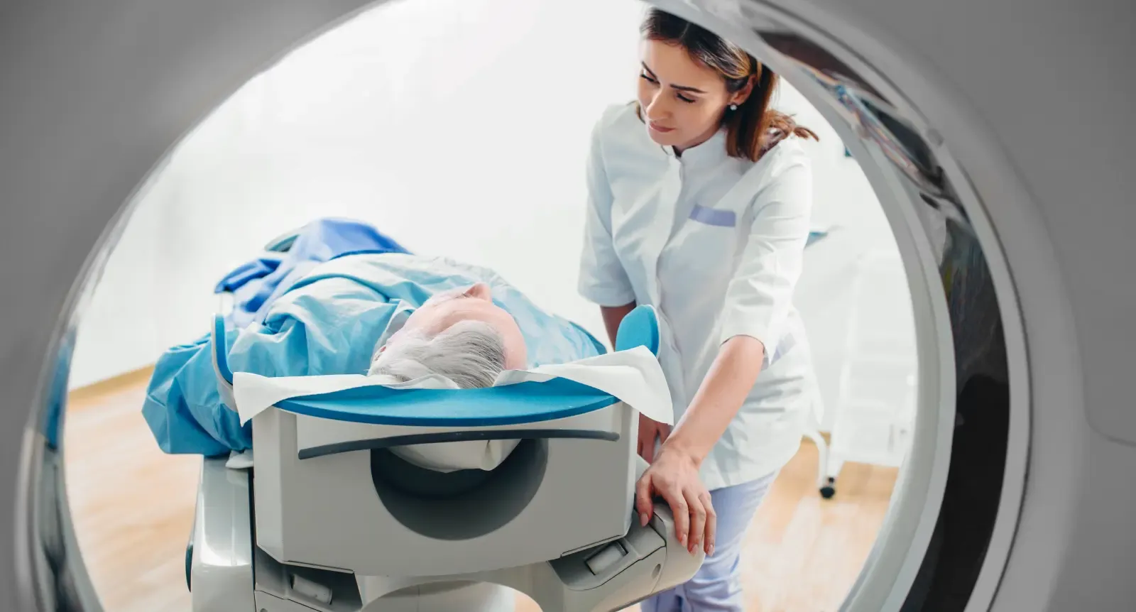
During your scan
Just before the scan starts, you may be given a medical dye called a contrast agent. This is given through a drip, usually into a vein in your hand or arm.
Your neuroradiographer will help you get comfortable as you lie in the scanner. Once you’re lying down, you’ll have a frame placed around your head called a head coil. They’ll leave the room while the scan is performed, but you can talk to them through an intercom.
The scanner moves around you and may make noises. It won’t touch you and you won’t feel it, though you may notice some tingling or warmth.
It’s important that you lie still during the scan to give accurate images. Our team will take every care to make you as comfortable as possible.
After your scan
You can usually go straight home after your MRI, unless you’re having other assessments.
Some people have mild side effects from the contrast agent, which can include feeling sick or having a headache. This usually passes quickly, and it helps to stay hydrated.
Your neuroradiologist will analyze your scans and send the results to your neurologist. This can take one to two weeks. Your neurologist will contact you when the results are ready. You'll have an appointment to discuss the results in person and plan any next steps.