PET-CT brain scan
What is a PET-CT brain scan?
A PET-CT scan is an advanced and sensitive imaging technology. It uses a small amount of a radioactive substance (called a radionuclide) to produce high spatial resolution images of brain structure and function. It’s used to diagnose several neurological conditions and track treatment progress.
It combines two scans:
- A PET scan, which uses a small amount of radiation to show brain activity
- A CT scan, which uses X-rays to show brain structures
When used together, these scans create a more complete picture of your brain health than when they’re used alone.
As a center of excellence in neurology, PET-CT imaging is part of our advanced diagnostic offering, helping to guide diagnosis and care planning for neurological conditions.


What is a PET-CT brain scan used for?
A PET-CT scan is a versatile imaging tool. You may have one on several occasions throughout your journey. Your neurologist may suggest a PET-CT scan to:
- Help diagnose a condition
- Track your condition’s progress
- Help design your treatment plan
- Monitor your response to treatment
PET-CT scans are commonly used to investigate conditions such as Alzheimer’s disease and other dementias, epilepsy, traumatic brain injuries (including chronic traumatic encephalopathy), and others.
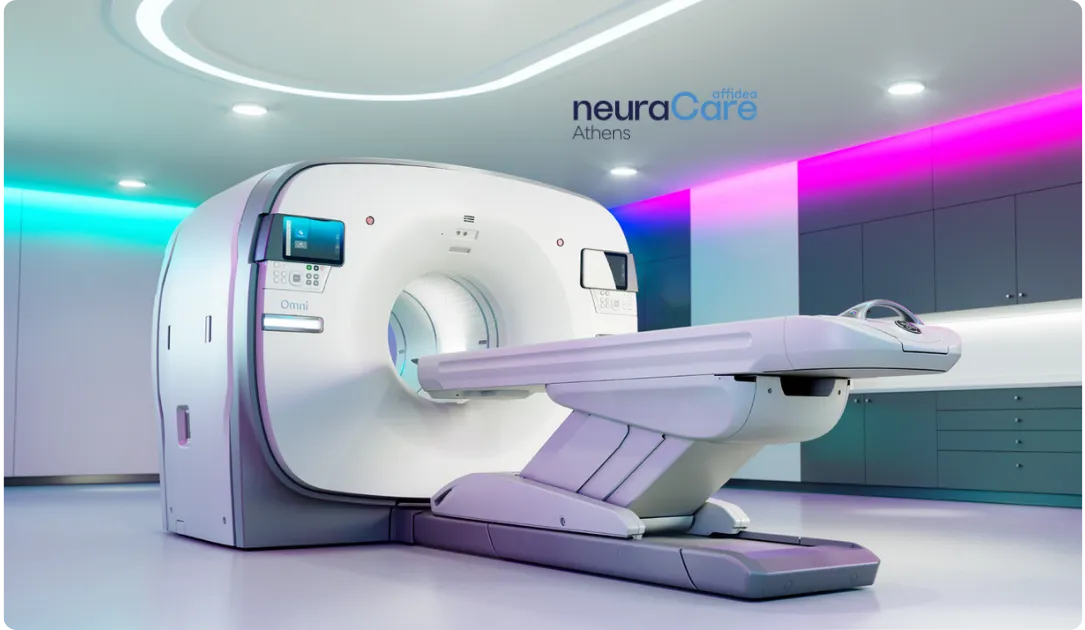
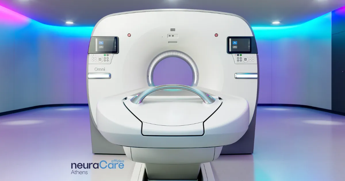
PET/CT OMNILegend 21 Technology at Affidea neuraCare
Affidea is the first in Greece, in collaboration with GE HealthCare, to introduce the technologically pioneering and internationally recognized award-winning PET/CT OMNI Legend 21cm 4R system. This system uniquely combines cutting-edge technologies and AI applications, making it the leading PET/CT system on the market, offering unparalleled benefits to patients while significantly enhancing the diagnostic confidence of the clinician:
- Superior detectability of small lesions/tumors
- Extremely low scan time and dose
- Best-in-class sensitivity
- Market-leading spatial resolution
- Advanced Respiratory Motion Management System
- Unique AI-based Image Reconstruction Algorithm with Precision DL
Advanced Digital Detection Design dBGO
The Omni Legend is based on GE HealthCare’s new digital dBGO detector – a unique combination of Digital Silicon Photomultipliers (SiPM) and small, highly sensitive dBGO crystals, which provide unmatched sensitivity and spatial resolution. The pioneering block architecture and advanced electronics deliver exceptional energy resolution and high count-rate performance, ensuring quantitative accuracy across all dose levels encountered in clinical practice.
The performance of the dBGO detector offers multiple advantages that place it at the forefront of modern systems, with the ability to detect very small lesions, enabling diagnosis in significantly shorter times and at lower doses compared to other systems available in the Greek market:
- Combines the excellent absorption capability of BGO with the detection power of SiPM
- Offers the industry’s largest axial field of view (AFOV)
- Provides outstanding spatial resolution (< 1.5 mm NEMA at 10 cm)
- Achieves excellent energy resolution (9.8%), allowing for narrow energy windows and resulting in outstanding scatter rejection (NEMA scatter fraction 35%)
Precision DL
At the core of the imaging functionality of the OMNI Legend system lies a unique advanced algorithm based on Precision DL neural networks, trained with thousands of Time-of-Flight (ToF) images. The combination of the exceptional sensitivity of the Omni Legend with Precision DL represents the future of PET/CT image quality.
The Omni Legend leverages innovations in artificial intelligence and deep learning, designed to deliver clearer images and greater diagnostic confidence. Precision DL is the most advanced solution for detecting small, low-contrast lesions compared to conventional PET/CT imaging systems.
Q.Clear
In today’s era of Precision Medicine, reliable quantitative assessment of radiopharmaceutical uptake in suspected lesions is a critical tool for diagnosis, staging, and monitoring of therapeutic response. Modern PET/CT systems use iterative image reconstruction algorithms, which are usually limited to 2 to 6 cycles, resulting in incomplete convergence of the raw data due to increasing noise. This often leads to underestimation of radiopharmaceutical uptake and, consequently, the extent of lesions.
The Q.Clear algorithm from GE HealthCare represents an innovative approach to full convergence, performing up to 25 iterations to confirm the image with reduced noise. The result is exceptionally accurate quantification of lesions and a high signal-to-noise ratio (SNR).
Beyond its accuracy in quantification and improved detection of small lesions, Q.Clear technology has been proven—according to international literature—to offer significant clinical benefits: 40% reduction in administered dose, 33% reduction in examination time, and a 62% improvement in SNR compared with ToF systems using conventional algorithms.
The OMNI Legend at Affidea neuraCare’s Centre of Excellence is equipped with the Q.Clear algorithm, offering in combination with Precision DL unparalleled accuracy, superior signal-to-noise ratio, maximum precision in lesion quantification, and therefore greater confidence in diagnosis and disease monitoring.
Auto-In
The correct and precise positioning of the patient in the system is a key factor for the success of an imaging examination. The position of the patient directly affects the quality of the images produced, the accuracy of the diagnosis, and the radiation dose received during the CT scan.
It is therefore crucial that the process is carried out accurately and quickly, ensuring both patient comfort and minimal radiation exposure for the operator.
The Auto-In function, with which our system is equipped, allows for contactless and fully automated patient positioning. This reduces the overall examination time, improves patient experience, and enhances staff safety.

Advanced Respiratory Motion Management System
GE HealthCare developed MotionFree, the first digital respiratory motion management technique without the use of external devices. MotionFree is designed to detect physiological respiratory motion in real time, dynamically determining whether the intensity of breathing creates imaging problems, and applying only the necessary corrections in the specific anatomical areas that require it.
MotionFree significantly improves the accuracy of quantitative analysis of radiopharmaceutical uptake in lesions that move during breathing and in determining the exact location and extent of lesions.
Dose Reduction Systems in CT Scans
Our system is equipped with the latest generation Q.AC algorithm from GE HealthCare, which uses ultra-low dose CT (ULD) data to perform highly accurate attenuation correction of PET images. With the Q.AC algorithm, attenuation correction is achieved with 10 times less radiation dose than CT scans in conventional systems.
Additionally, the system includes the latest-generation CT iterative reconstruction algorithm, ASiR-V, contributing to:
- Reduced CT dose: Dose reduction up to 50%–82% compared to the classic FBP algorithm, while maintaining stable dose levels during the scan.
- Improved lesion contrast sensitivity: Contrast improvement of 59%–133% while maintaining stable dose levels during the scan.
- Reduced image noise: Noise reduction of up to 56% and 91% depending on dose levels during the scan.
- Improved spatial resolution: Up to 2.07% (107%) improvement while maintaining stable dose levels during the scan.
At the same time, the ASiR-V algorithm has been shown to reduce false positives compared to FBP, further increasing diagnostic confidence.
Book a consultation
How to prepare for a PET-CT brain scan
Before your appointment, your neurologist may ask you to:
- Not eat or drink anything other than water for six hours
- Remove anything with metal before your scan, such as jewelry, wired undergarments, or belts
- Avoid intense physical activity, such as running, cycling, and going to the gym
If you need to wear metal, such as hearing aids or glasses, that’s okay. You can wear them to your appointment and remove them before your scans.
Your neurologist will also speak to you in advance if:
- You’re pregnant or breastfeeding
- You take any medication
- You’re diabetic

Our multidisciplinary team
We surround our patients with specialists who listen, understand, and care. They include some of Europe's most renowned experts – neurologists,
neuropsychologists, neuroradiographers, microbiologists, nurses, nuclear medicine experts and therapeutic specialists in physiology and occupational health.
What to expect during a PET-CT brain scan
An experienced neuroradiologist performs your PET-CT scan. This is a doctor who specializes in brain and spinal cord imaging. They’ll guide you throughout your appointment and help you understand what to expect.
Before the scan
Your neuroradiologist will ask you to remove any items containing metal, such as jewelry, hearing aids, or glasses. They may ask you to change into a hospital gown for your comfort.
An hour before your scan, you’ll have the radiotracer injected. This is usually into a vein in your arm or hand through a cannula (a small plastic tube).
The amount of radiation is very low and does not make you radioactive. It will leave your body after a few hours, and side effects are extremely rare. If you have any concerns, your expert neuroradiologist is there to help you.
During the scan
A PET-CT scanner is a large machine with a hole in the center and a bed attached to it. Your scans are taken inside the hole of the scanner and can take between 30 to 60 minutes.
- Your neuroradiologist will help you into the correct position on the bed. They’ll help make sure you’re comfortable.
- They will leave the room to take the scan. You’ll still be able to communicate with each other, and they’ll be able to see you from the other room.
- The bed will slowly move in and out of the hole as it takes pictures. It’s important to lie still and follow any instructions your neuroradiologist gives.
If you’re worried about lying still or being in an enclosed space, let your neuroradiologist know. We can help make sure you’re as comfortable and relaxed as possible.
After the scan
If you don’t have any other tests or treatments, you’re free to go home straight after your appointment.
Before you leave, your neuroradiologist may give you some safety tips to follow, such as:
- To drink plenty of water to help flush the radionuclide from your body
- To avoid prolonged or close contact with young children and pregnant women for the rest of the day
Your neuroradiologist will analyze your scans and send the results to your neurologist. This can take 1 to 2 weeks . Your neurologist will contact you when the results are ready. You'll have an appointment to discuss the results in person and plan any next steps