SPECT-CT scan

What is a SPECT-CT scan?
A SPECT-CT scan is an important imaging technique in neurology. It produces images of your brain’s inner structures and activity by combining two different scans:
- A SPECT scan, which uses a radioactive substance (called a radionuclide) that produces gamma rays to show areas in the brain with high activity
- A CT scan, which uses X-rays to create a map of your brain structures
When these scans are combined, we can more accurately identify areas in the brain that are not working as well as they should be. This helps neurologists diagnose, monitor, and plan treatment for several neurological conditions.
As a center of excellence in neurology, we use SPECT-CT as one of several up-to-date technologies for diagnosing neurological conditions in our world-class diagnostic suites, designed for patient comfort and fast, accurate imaging.

What’s a SPECT-CT scan used for?
Many people with neurological conditions will have brain imaging scans at several stages of their journey. You may have a SPECT-CT scan to help:
- Diagnose your condition
- Monitor your condition’s progression
- Design your personalized treatment plan
- Assess your treatment response
Common conditions we use a SPECT-CT scan to investigate include seizures and epilepsy, dementia, headaches and migraines, and traumatic brain injuries.
Book a consultation
The SPECT-CT technology at Affidea neuraCare
SPECT/CTNM/CT 850
Affidea neuraCare is the first centre of excellence in Greece to be equipped with the most advanced hybrid SPECT/CT NM/CT 850 system in collaboration with GE HealthCare. Staying true to our commitment to innovation, we have equipped our centre with this cutting-edge SPECT/CT system, specially designed for the new era of Theranostics and Precision Medicine.
The NM/CT 850 from GE HealthCare integrates the latest state-of-the-art technologies, delivering outstanding diagnostic accuracy. It strengthens physicians’ confidence thanks to superior image quality, while at the same time ensuring minimal radiation exposure and impressively fast acquisition times.
All these elements contribute to an absolutely safe and effective experience for our patients. The system includes a suite of advanced technologies that guarantee accuracy in diagnostic outcomes with exceptionally low dose levels for the patient.

Pioneering Application of LEHRS Collimators (Low Energy High Resolution Sensitivity)
The NM/CT 850 system is the first SPECT/CT from GE HealthCare in Greece to be equipped with the most advanced LEHRS photon collimator, which provides even greater sensitivity in radiation detection compared to conventional systems. As a result, it significantly reduces the overall scan time and the radiation dose for the patient.
The pioneering design of LEHRS is part of the state-of-the-art SwiftScan technology chain, which allows the system to capture up to 20% more photons during the scan. This leads to reduced acquisition times and up to 25% lower administered doses compared to conventional SPECT/CT systems.
This innovative technology significantly improves the detectability of small lesions without additional time or dose, enhancing imaging of lesions and metastases at early and critical stages. It is available across all examinations in conventional Nuclear Medicine.
Evolution
The NM/CT system comes equipped with the pioneering Evolution for Bone, Evolution for Planar Bone, and Evolution Toolkit programs. These are a set of tools that monitor the operation and communication between the collimator-detector used in the system’s head and the detector arrangement, then process and correct the parameters that could affect the clinical outcome.
The Evolution for Bone algorithm, developed at Johns Hopkins University and UNC Chapel Hill, models the system’s collimator-detector response, improves image analysis and signal-to-noise ratio, and significantly reduces noise variability.
The use of this option allows:
- Imaging in half the time compared to conventional system protocols with typical dose administration
- Imaging with half the dose compared to conventional system protocols with standard acquisition time
Overall, the Evolution program, in combination with SwiftScan technology, can reduce examination time or administered dose by up to 75%, providing a safer and more efficient experience for the patient.
How to prepare for a SPECT-CT scan
Generally, you do not need to do anything differently before your scan, meaning you can eat and drink as usual. Your neurologist will speak to you if you:
- Are pregnant or breastfeeding
- Are taking any medication
It’s a good idea to avoid wearing items with metal, such as jewelry, clothes with zips or wire, or belts. Some items with metal are fine to wear to your appointment, such as hearing aids and glasses. You can remove them before your scan.

Our multidisciplinary team
We surround our patients with specialists who listen, understand, and care. They include some of Europe's most renowned experts – neurologists,
neuropsychologists, neuroradiographers, microbiologists, nurses, nuclear medicine experts and therapeutic specialists in physiology and occupational health.
What to expect during a SPECT-CT scan
A neuroradiologist takes your scan. This is a doctor who specializes in brain imaging. They’ll help explain what to expect and answer any concerns.
Before your scan
If you’re wearing anything with metal, your neuroradiologist will ask you to remove it before the scan begins. They may ask you to change into a gown for your comfort.
Before your SPECT-CT scan, your neuroradiologist will inject a radiotracer into a vein in your arm or hand. It’s usually injected through a cannula, which is a small plastic tube.
The tracer contains a small amount of radiation, which allows us to see how blood is flowing in the brain. It will not make you radioactive or cause any side effects, and it will leave your body after a few hours. We can answer any questions you have about this.
During your scan
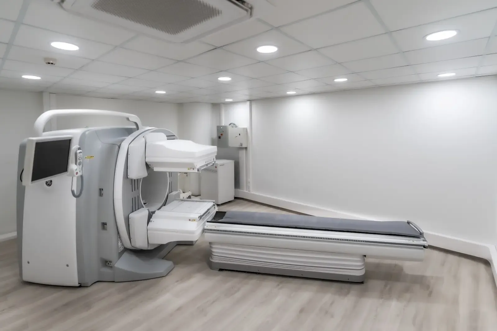
A SPECT-CT scanner is a large machine with a hole in the center and a flat couch attached. The camera moves around you during the scan, which can take up to 40 minutes.
Both scans are performed on the same machine, one after the other.
- Your neuroradiologist will help position you on the couch and make sure you’re comfortable.
- They’ll go into the room next door to take the scans. They can still see you and you can talk to each other through the speaker system.
- When the scan begins, the couch will move in and out of the machine as the camera moves around you. It’s important to lie very still and listen to any instructions from your neuroradiologist.
If you’re concerned about lying still or being inside an enclosed space, let your neuroradiologist know. We’re here to help you be as comfortable as possible.
After your scan
You can go straight home after your scan if you don’t have any other tests, scans or appointments. Your neuroradiologist will discuss some safety advice before you go, such as:
- Drinking plenty of fluids to help flush the radionuclide from your body
- Avoiding prolonged close contact with small children and pregnant women for a few hours after your scan
Your neuroradiologist will carefully analyze your scans and share the results with your neurologist . This can take up to 2 weeks. Your neurologist will schedule an appointment to discuss your results in person and any next steps.